After a number of attempts to use the White Light Profilometer, which didn’t come off for various reasons, I instead made arrangement to use the microscopes. PhD candidate Tony Scimone supported me to use the equipment and navigate the culture of the lab (I wore a lab coat, which he apologised for but I felt strangely performative and absorbed into the environment).
The microscopes we used are optomised for flat/2D samples (slides) but the objects I brought had more depth, so he was unsure what the results we might get.
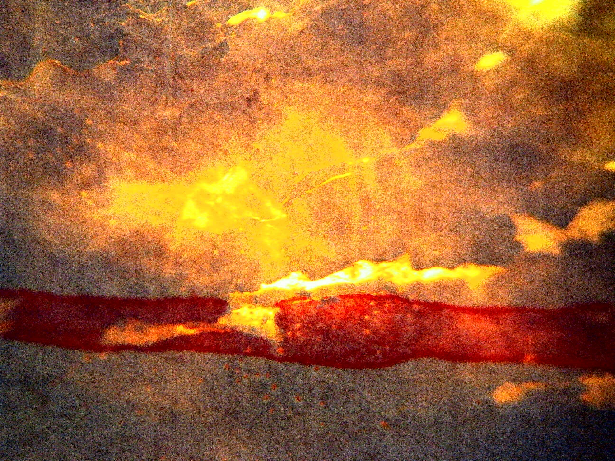
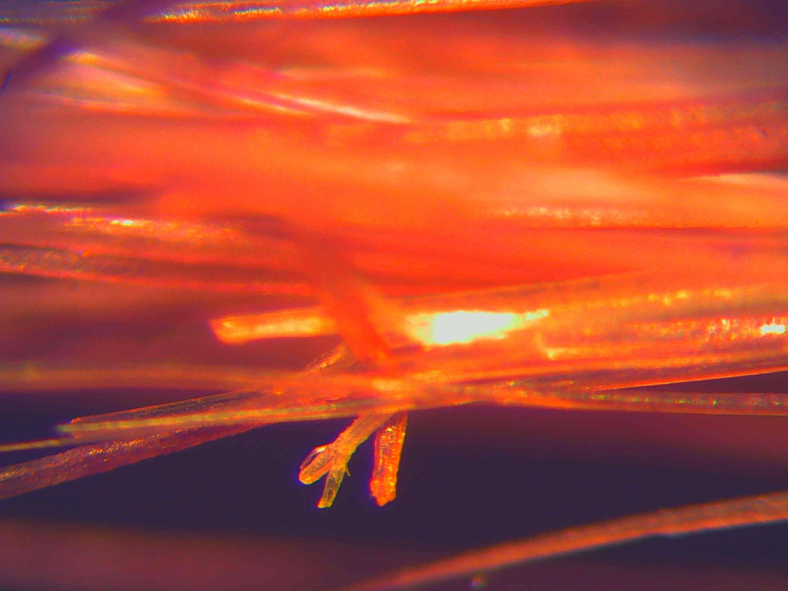
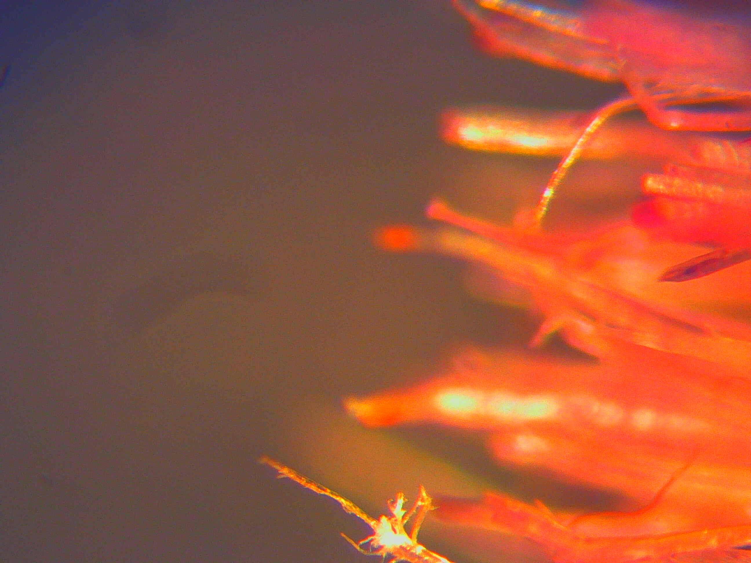
The first microscope we used was a Bright Field microscope, which means the sample is lit from the bottom.
We enlarged the image between 40x and 100x. The above image and one below are from a scrap of wallpaper from the wall of my studio, which is painted magnolia and has a splatter of red paint on it:
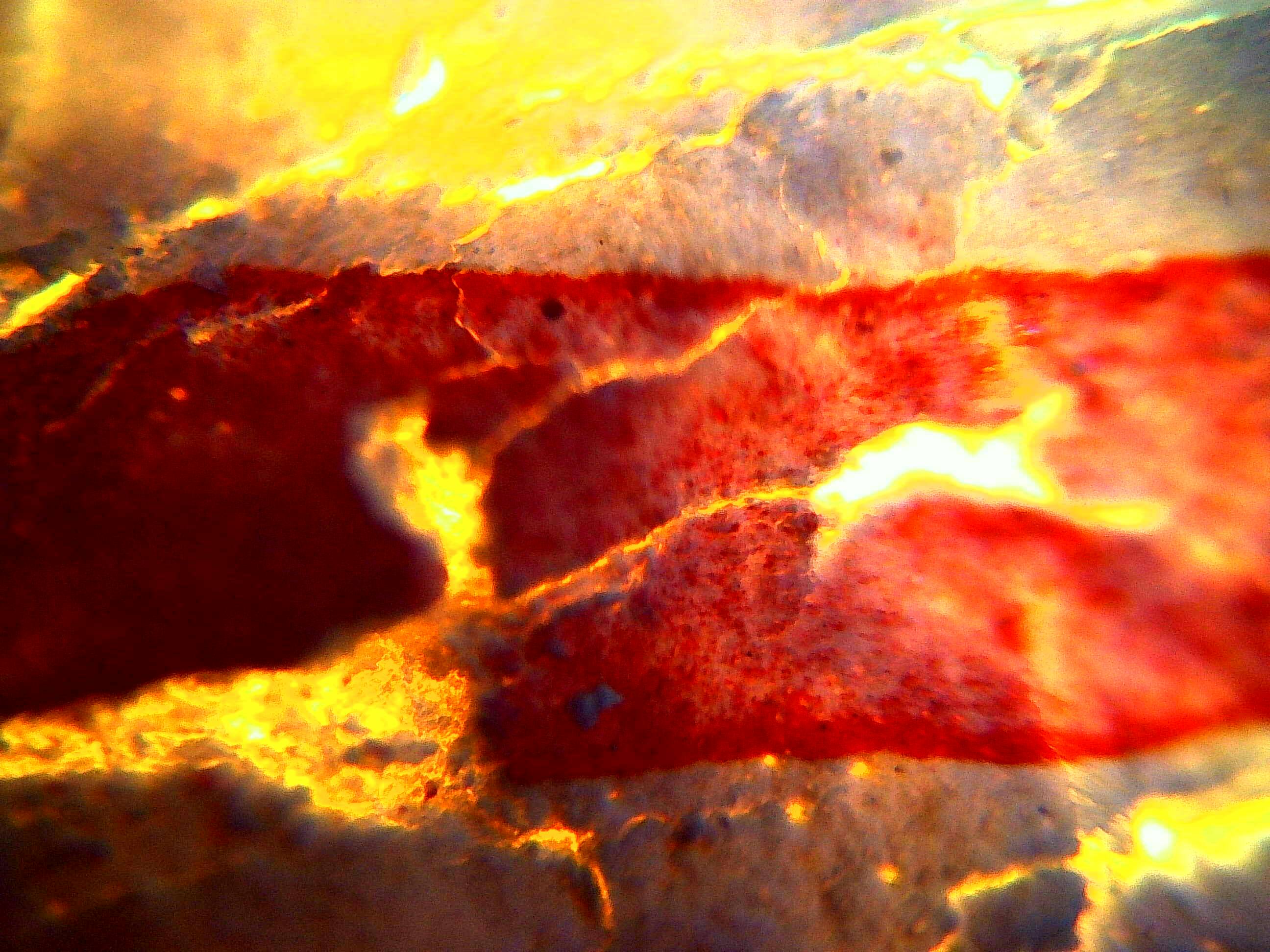
These images remind me of a volcanic landscape or something from Lord of the Rings. Scimone said it reminded him of the cover of Metallica’s album ‘Reload’ (looking at it, I think my images are far more interesting!) You can see from the depth of field that the splatter of paint is raised above the paper.
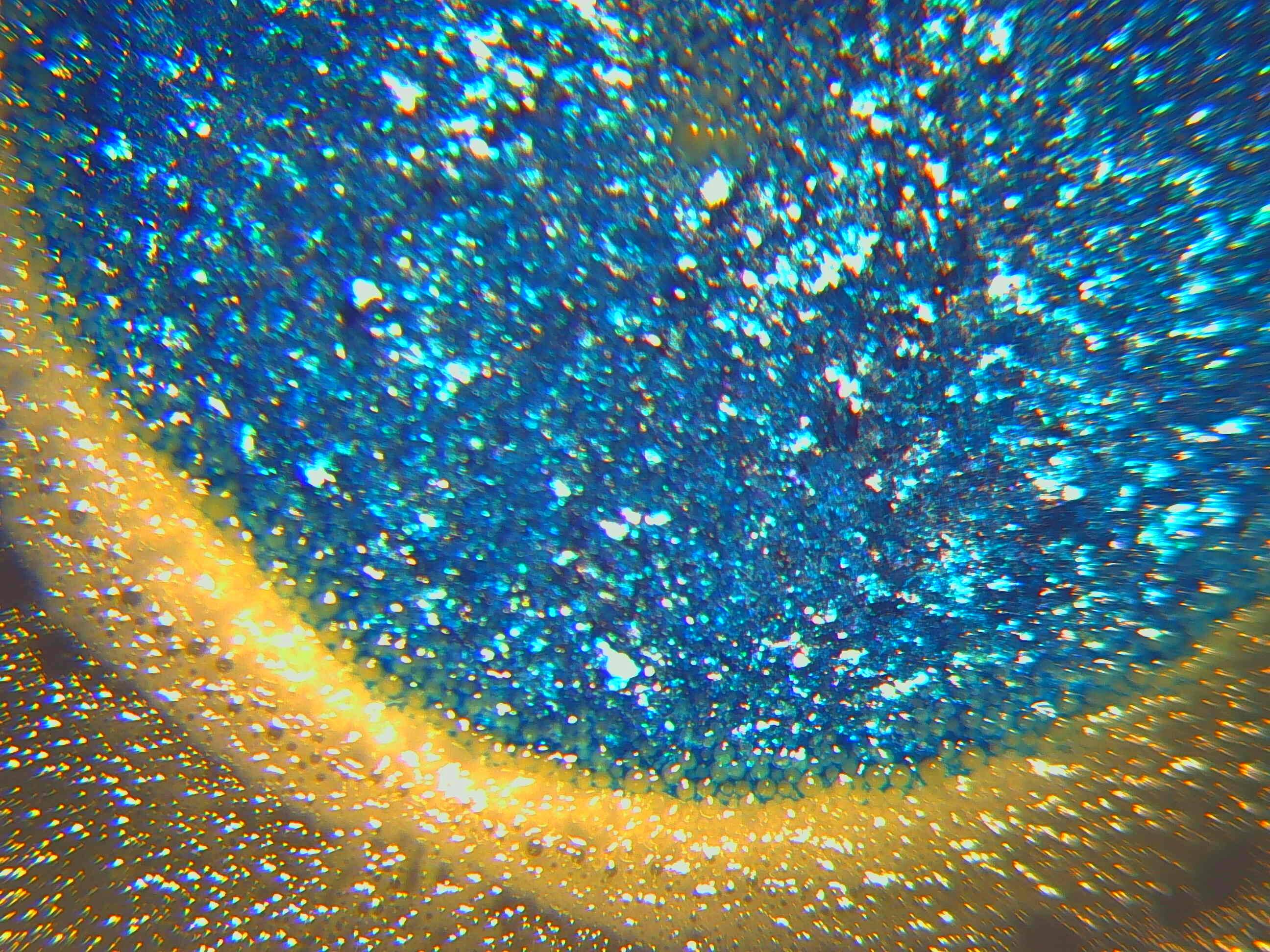
For the following images we used a ‘Top Down’ microscope, where the light comes form above the sample rather than below. These images are from a sample of blue tac taken from the wall of the studio:
At this level, you can see hairs and textures that are not visible to the naked eye. There are also small particles of wall and other debris which have come away with the blue tac. When trying to capture this image, we noticed that it looked like it was moving on the digital screen. This was because the heat from the light and the malleability of the material meant there was some slight movement, which was totally not visible to the naked eye. As I watched Scimone move the sample under the lens, it was interesting to note how it zoomed around on the digital screen when he was moving it fractions of a millimeter. Clearly there is a craft to microscopy that can be honed over years of practice!
The final object I had brought from the studio was a paint brush. We photographed the bristles:
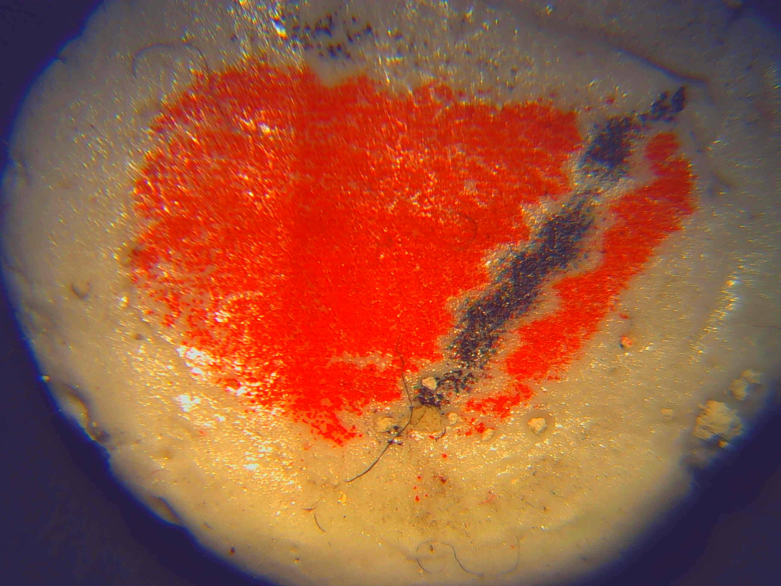
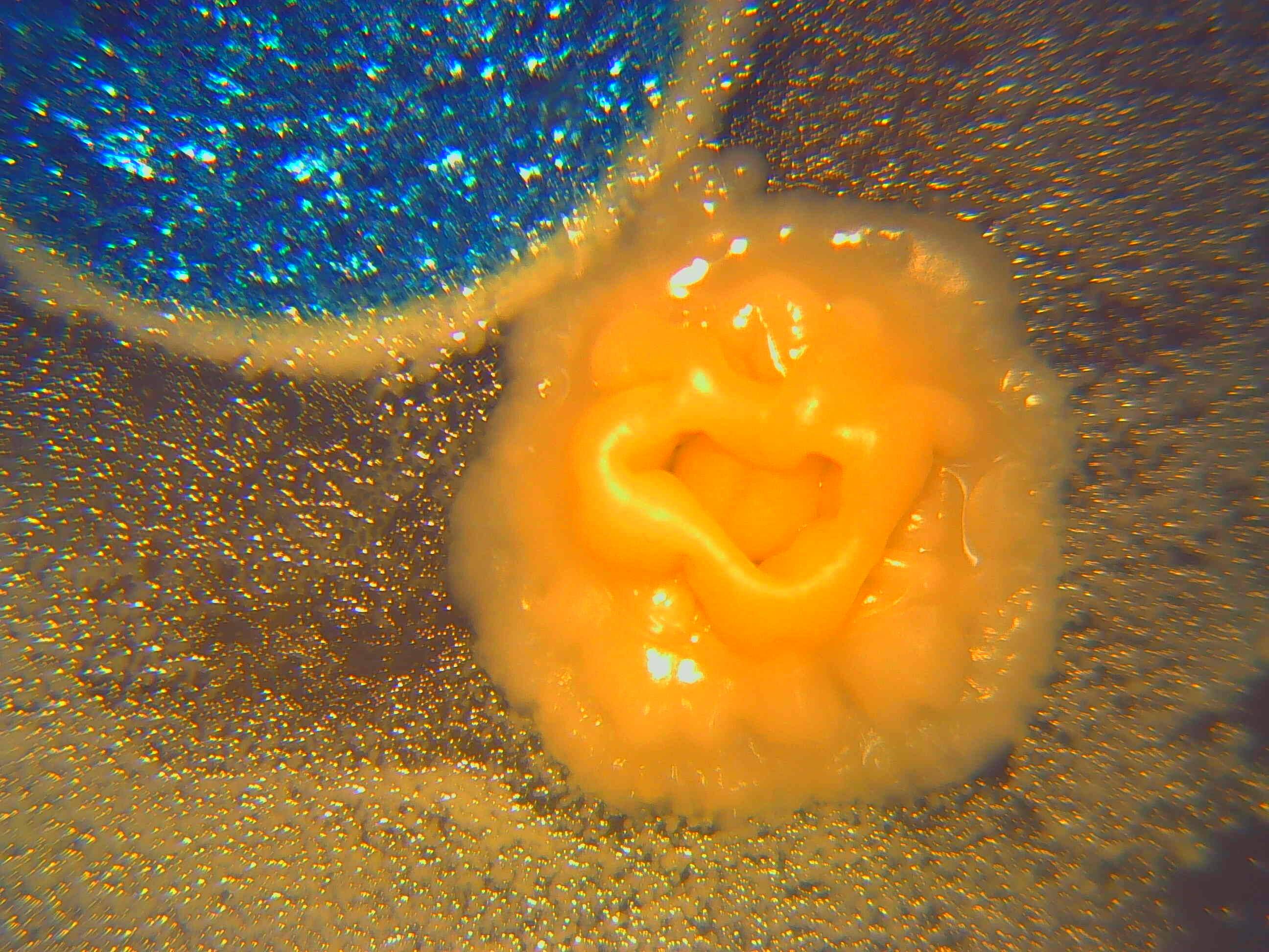
As well as the objects from the studio, I had a test plate with me that had produced interesting results, so we also looked at that under the microscope. During taking these images, we had a Bunsen burner lit nearby. This is to create an area of hot air and convection around us that would prevent airborne microbes that could contaminate the samples.
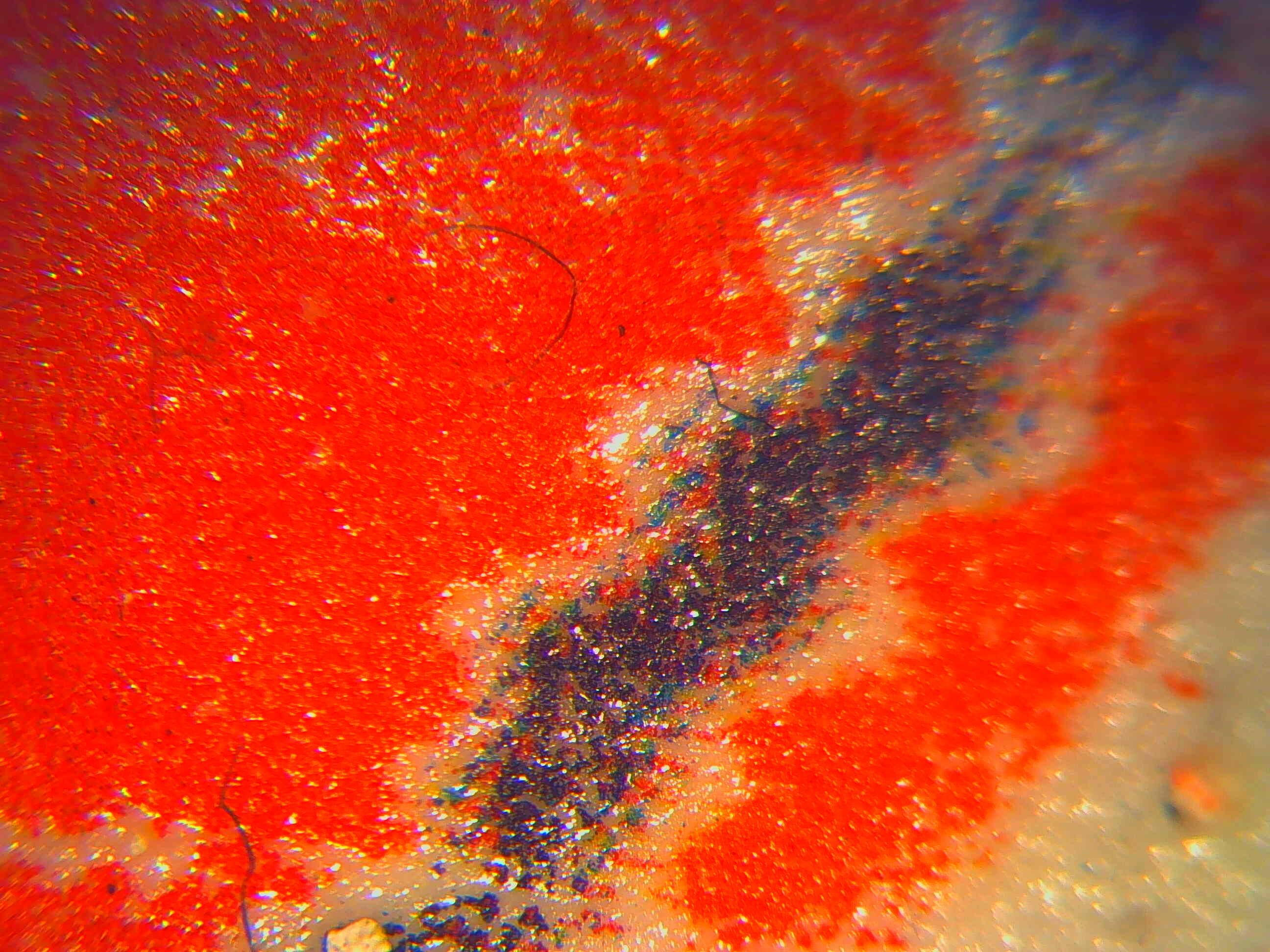
The above image shows how the cultures join up to the paint spot, as well as a small area of microbes that have grown directly in the middle of the paint. The below image shows a different culture of microbes on the same plate:
Scimone was a real expert with the technology so the most difficult thing was getting the images files from the device so I could take them home! There are many more images than included in this blog that I haven’t got access to myself yet.
While we were taking these photos, I took the opportunity to ask Scimone some questions about his work and the life of a microbiologist.
One of the misconceptions that he comes up against frequently is the idea that all bacteria = bugs = disease, or, that viruses and bacteria are the same thing, which is radically untrue. The scale between viruses and bacteria is the same as the scale between bacteria and ourselves (viruses are much, much smaller), and there are some viruses which only infect bacteria.
A favourite factoid of Scimone’s that he likes to share on university open days is that the bacteria Actinomyces evolved in the same way as fungi – in that they both form hyphae and mycelium, and have spores – but this evolution happened millions of years apart and totally independently of each other, on an entirely different branch of the tree of life.
Considering this rather specific growth and lifecycle, it’s this evolutionary distance that I find fascinating – they’re in different kingdoms of life altogether, so the idea that hyphae and mycelia were evolved independently millions of years apart really excites me!
He also commented that there are no such thing as ‘friendly’ bacteria, as often characterised in certain foodstuff adverts.Bacteria are either pathogenic, non-pathogenic, or opportunistically pathogenic. In fact, some of the bacteria that live in our gut could kill us if they got into our bloodstream.
We also talked briefly about the philosophy of science and in particular the idea of subjectivity as an influence on the scientific method. This is something I’m interested in, and plan to look at the work of Karen Barad later to explore this further. Although this isn’t a major concern of the MMU education provision, it seem that there is some interest in this in the faculty and some recognition of the agency and influence of the researcher in experiments. Certainly this is an area that opens up fruitful dialogue with the School of Art and other disciplines. I’ve enjoyed seeing how similar some of our concerns are when you strip them down to the bare essentials: authenticity, process, reality, application. Scimore did explain how scientists seek to minimise subjectivity:
Researchers address subjectivity in their work through appropriate statistical tests. We ourselves as researchers always have an element of bias and subjectivity, but by collecting data and allowing independent mathematical processes to handle the data once we’ve compiled it, we can validate scientific claims from an objective point of view (at least more objective).
I asked whether the images we had produced here showed us something of the world from a microbes’ perspectives. He replied that the general rule is you need a 1000x magnification, which we hadn’t gone up to (because the samples were not flat enough).
Despite this, I am really happy with these images. I didn’t have any idea what to expect from this session, and I feel the results have huge aesthetic quality, appearing as landscapes with depth and contrast. I’m not yet sure how I will use these in my final presentation, though my feeling is I will blow them up and produce vinyl to go on glass doors in the exhibition space, further enhancing their transparent quality and the effect of light passing though them. This could also make the viewer feel momentarily tiny themselves, like the microbes that live on these precarious planes.
References:
Scimone, T. (2017) Interview/conversation/workshop event at MMU, 02/02/17.